
MRI Test
- Home
- MRI Test
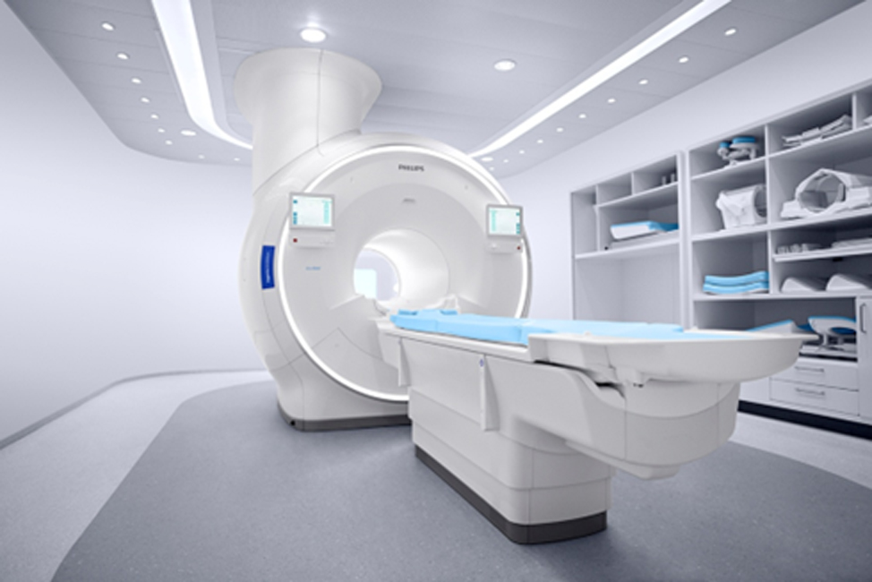
What is 3 tesla MRI?
Scanner consists of a large and very strong magnet in which the patient lies. A radio wave antenna is used to send signals to the body and then receive signals back. These returning signals are converted into pictures by a computer attached to the scanner. MRI is quite safe in the majority of patients. Certain patients may not be able to have an MRI.
MRI scan facts
- MRI scanning uses magnetism, radio waves, and a computer to produce images of body structures.
- Scanning is painless and does not involve x-ray radiation.
- Patients with heart pacemakers, metal implants, or metal chips or clips in or around the eyes cannot be scanned with MRI because of the effect of the magnet.
- Claustrophobic sensation can occur with MRI scanning.
During the test
The MRI machine looks like a tube that has both ends open. You lie down on a movable table that slides into the opening of the tube. A technologist monitors you from another room. You can talk with the person by microphone.
Machine creates a strong magnetic field around you, and radio waves are directed at your body. The procedure is painless. You don’t feel the magnetic field or radio waves, and there are no moving parts around you.
During the MRI scan, the internal part of the magnet produces repetitive tapping, thumping and other noises. Earplugs or music may be provided to help block the noise. If you are worried about feeling claustrophobic inside the MRI machine, talk to your doctor beforehand. You may receive a sedative before the scan.
In some cases, a contrast material, typically gadolinium, may be injected through an intravenous (IV) line into a vein in your hand or arm. The contrast material enhances the appearance of certain details. The material used for MRIs is less likely to cause an allergic reaction than the material used for CT scans.
An MR can last up to an hour or more. You must hold very still because movement can blur the resulting images.
During a functional MRI, you may be asked to perform a number of small tasks — such as tapping your thumb against your fingers, rubbing a block of sandpaper or answering simple questions. This helps pinpoint the portions of your brain that control these actions.
What is CT SCAN?
CT scanning—sometimes called CAT scanning—is a noninvasive medical test that helps physicians diagnose and treat medical conditions. CT scanning combines special x-ray equipment with sophisticated computers to produce multiple images or pictures of the inside of the body. These cross-sectional images of the area being studied can then be examined on a computer monitor, printed or transferred to a CD.
CT scans of internal organs, bones, soft tissue and blood vessels provide greater clarity and reveal more details than regular x-ray exams.
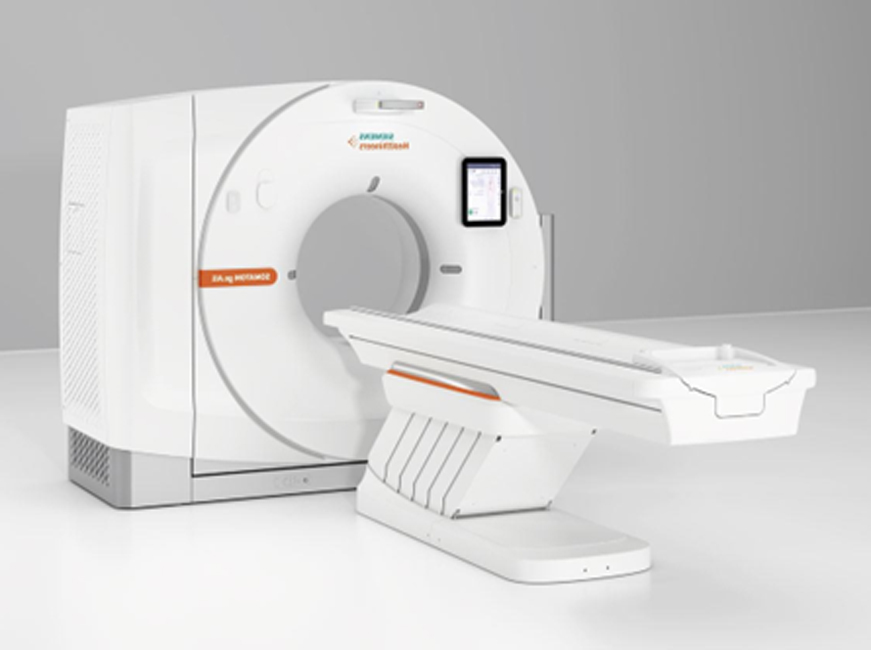
How is a CT scan done?
CT stands for computerised tomography, and CAT for computerised axial tomography (sometimes the word computed is used instead of computerised). The CT scanner looks like a giant thick ring. Within the wall of the scanner there is an X-ray source. Opposite the X-ray source, on the other side of the ring, are X-ray detectors. You lie on a couch which slides into the centre of the ring until the part of the body to be scanned is within the ring. The X-ray machine within the ring rotates around your body. As it rotates around, the X-ray machine emits thin beams of X-rays through your body, which are detected by the X-ray detectors.
What is a CT scan used for?
A CT scan can be done on any section of the head or body. It can give clear pictures of bones. It also gives clear pictures of soft tissues which an ordinary X-ray test cannot show, such as muscles, organs, large blood vessels, the brain and nerves. The most commonly performed CT scan is of the brain – to determine the cause of a stroke, or to assess serious head injuries. Other uses of a CT scan include:
To detect abnormalities in the body, such as tumours, abscesses, abnormal blood vessels, etc, when they are suspected by symptoms or other tests.
To give a surgeon a clear picture of an area of your body before certain types of surgery.
To pinpoint the exact site of tumours prior to radiotherapy.
To help doctors find the right place to take tissue samples (biopsies).
CT –SCAN PRICE LIST
| S.NO | TYPE OF STUDY | PLAIN STUDY | CONTRAST STUDY | ||||
| 1 | CT BRAIN | 2500 | 3500 | ||||
| 2 | CT SELLA | 2500 | 3500 | ||||
| 3 | CT CVJ | 4000 | 5000 | ||||
| 4 | CT ORBIT (AXIAL AND CORONAL) | 4500 | 5500 | ||||
| 5 | CT PNS (CORONAL) | 4000 | 5000 | ||||
| 6 | CT PNS (AXIAL AND CORONAL) | 4500 | 5500 | ||||
| 7 | CT FACE | 4500 | 5500 | ||||
| 8 | HRCT TEMPORAL BONE | 5000 | 6000 | ||||
| 9 | CT HEAD + ORBIT | 4500 | 5500 | ||||
| 10 | CT HEAD + ORBIT + PNS | 5500 | 6500 | ||||
| 11 | CT NECK | 4500 | 5500 | ||||
| 12 | CT CHEST | 4500 | 6000 | ||||
| 13 | HRCT CHEST | 5000 | 6000 | ||||
| 14 | CT CHEST + HRCT | 5500 | 6500 | ||||
| 15 | CT FACE + NECK | 5500 | 6500 | ||||
| 16 | CT NECK + CHEST | 6500 | 8000 | ||||
| 17 | CT NECK + UPPER CHEST | 5500 | 6500 | ||||
| 18 | CT UPPER ABDOMEN | 4500 | 6000 | ||||
| 19 | CT LOWER ABDOMEN | 4500 | 6000 | ||||
| 20 | CT WHOLE ABDOMEN | 6000 | 9000 | ||||
| 21 | CT CHEST & UPPER ABDOMEN | 6000 | 8500 | ||||
| 22 | CT ABDOMEN DUAL PHASE | 10,500 | |||||
| 23 | CT ABDOMEN TRIPLE PHASE | 11,500 | |||||
| 24 | CT NECK + CHEST + ABDOMEN | 9500 | 12,000 | ||||
| 25 | CT NECK + CHEST + UPPER ABDOMEN | 8000 | 11,500 | ||||
| 26 | CT CHEST + ABDOMEN | 9000 | 11000 | ||||
| 27 | NCCT KUB | 5000 | |||||
| 28 | CECT KUB | 7000 | |||||
| 29 | CT UROGRAPHY | 10,500 | |||||
| 30 | CT UROGRAM + CT ABDOMEN | 8000 | 12,500 | ||||
| 31 | CT ENTEROGRAPHY | 10,500 | |||||
| 32 | CT RENAL ANGIO + CT UROGRAM | 13,500 | |||||
| 33 | CT WHOLE SPINE | 13,500 | 15000 | ||||
| 34 | CT SPINE (ONE LEVEL) | 4500 | 5500 | ||||
| 35 | CT SPINE (EXTRA ONE LEVEL) SCREEN | 2000 | |||||
| 36 | CT JOINT / EXTREMITY | 4500 | 5500 | ||||
| 38 | CT 3D RECONSTRUCTION PER SITE | 2000 | |||||
| 39 | CT CHEST +ENTEROGRAPHY | 13,500 | |||||
| 40 | CT VIRTUAL BRONCHOSCOPY | 9500 | |||||
| 41 | CT VIRTUAL COLONOSCOPY | 10,500 | |||||
| 42 | CT AORTOGRAM / CT PULMONARY ANGIO | 14,000 | |||||
| 44 | CT DUAL PHASE ABDOMEN & VOLUMENTRY LIVER | 12000 | |||||
| 45 | CT ANGIOGRAPHY BRAIN /CAROTID/RENAL | 11,000 | |||||
| 46 | CT BRAIN + NECK ANGIOGRAPHY | 15,000 | |||||
| 47 | CT ANGIOGRAPHY UPPER / LOWER LIMBS (2) | 12,000 | |||||
| 48 | CT ANGIOGRAPHY ABDO + LOWER LIMBS | 16,000 | |||||
| 49 | CT ANGIOGRAPHY ABDOMEN | 11000 | |||||
| 50 | CT CORONARY ANGIOGRAPHY | 15,500 | |||||
| 51 | CT CORONARY CALCIUM SCORE | 8000 | |||||
| 52 | CT SINOGRAM | 6000 | |||||
3.Tesla M.R.I PRICE LIST
| TYPE OF STUDY | PLAIN STUDY | CONTRAST STUDY | |
| 1 | MRI BRAIN | 8000 | 11000 |
| 2 | MRI BRAIN WITH DIFFUSION ( STROKE PROTOCOL) | 11000 | 14000 |
| 3 | MRI BRAIN WITH PERFUSION | 11000 | 14000 |
| 4 | MRIN BRAIN WITH EPILEPSY PROTOCOL | 11000 | 14000 |
| 5 | MRI BRAIN WITH SPECTROSCOPY | 16000 | 19000 |
| 6 | MRI BRAIN WITH ANGIOGRAPHY | 16000 | 19000 |
| 7 | MRI BRAIN WITH VENOGRAPHY | 16000 | 19000 |
| 8 | MRI BRAIN WITH INNER EAR (3D COCHLEA) | 11000 | 14000 |
| 9 | MRI SPECTROSCOPY | 8000 | |
| 10 | MRI BRAIN WITH CSF FLOW STUDY | 12000 | 15000 |
| 11 | MRI BRAIN WITH CISTERNOGRAPHY | 18000 | 21000 |
| 12 | MRI CISTERNOGRAPAHY | 11000 | 14000 |
| 13 | MRI ORBIT | 8000 | 11000 |
| 14 | MRI BRAIN WITH ORBIT | 11000 | 14000 |
| 15 | MRI FACE WITH NECK | 8000 | 11000 |
| 16 | MRI VENOGRAPHY BRAIN | 8000 | 11000 |
| 17 | MRI ANGIOGRAPHY BRAIN | 8000 | 11000 |
| 18 | MRI ANGIOGRAPHY BRAIN + NECK | 16000 | 19000 |
| 19 | MRI CV JUNCTION | 8000 | 11000 |
| 20 | MRI BRAIN WITH PITUITARY/ SELLA CUTS | 11000 | 14000 |
| 21 | MRI DYNAMIC CONTRST STUDY PITUITARY | 15000 | |
| 22 | MRI PARANSAL SINUS / MRI FACE | 8000 | 11000 |
| 23 | MRI NECK | 8000 | 11000 |
| 24 | MRI ANGIOGRAPHY NECK VESSELS | 8000 | 11000 |
| 25 | MRI CHEST | 9000 | 12000 |
| 26 | MRCP | 8000 | 11000 |
| 27 | MRI UPPER / LOWER ABDOMEN | 8000 | 11000 |
| 28 | MRI UPPER ABDOMEN + MRCP | 11000 | 14000 |
| 29 | MRI WHOLE ABDOMEN | 16000 | 19000 |
| 30 | MRI TRIPPLE PHASE LIVER/ DYNAMIC PANCREAS | 15000 | |
| 31 | MRI ENTEROGRAPHY | 16000 | 19000 |
| 32 | MRI UROGRAPHY | 11000 | 14000 |
| 33 | MRI UPPER ABDOMEN + UROGRAPHY | 19000 | 22000 |
| 34 | MRI RENAL ANGIOGRAPHY | 10000 | 13000 |
| 35 | MRI PELVIS | 8000 | 11000 |
| 36 | MRI PELVIS WITH FIBROID MAPPING | 11000 | 14000 |
| 37 | MRI PROSTATE ( MULTIPARA DYNAMIC) | 15000 | |
| 38 | MRI FISTULOGRAM/SINOGRAM | 8000 | 11000 |
| 39 | MRI PELVIC FLOOR DYNAMIC STUDY | 15000 | |
| 40 | MRI SPINE PART (CERVICAL / DORSAL / LUMBAR) | 8000 | 11000 |
| 41 | MRI WHOLE SPINE SCREEENING | 9000 | |
| 42 | MRI WHOLE SPINE | 24000 | 27000 |
| 43 | MRI BOTH SI JOINT | 8000 | 11000 |
| 44 | MRI JOINT | 8000 | 11000 |
| 46 | MRI JOINT WITH CARTIGRAM | 12000 | 15000 |
| 47 | MRI JOINT WITH ARTHROGRAM | 20000 | |
| 48 | MRI BRACHIAL PLEXUS | 8000 | |
| 49 | MRI FULL LIMB ( ARM / LEG) | 16000 | 19000 |
| 50 | MRI ANGIOGRPAHY BOTH LIMBS | 19000 | |
| 51 | MRI VENOGRAPHY BOTH LIMBS | 19000 | |
| 52 | MRI (BREAST) MAMMOGRAPHY | 15000 | |
| 53 | CARDIAC MRI | 18,500 | |
| 54 | MRI ANTENATAL | 10000 |